Endoscopic Ultrasound: Understanding your Heath
EUS allows a physician to get a clear picture of organs and tissues using a tool that combines an endoscope with a tiny ultrasound unit. Ultrasound projects sound waves into body tissues and then “listens” as the sound waves “echo” back to a sensor. The sensor creates a visual image on a screen that can be evaluated by the physician.
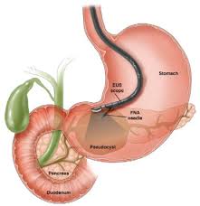
In EUS, the ultrasound probe is built onto the end of an endoscope-a lighted, flexible rube that can be fed through the mouth and into the esophagus (the tube that carries food to the stomach) or through the rectum, depending on what part of the digestive tract the physician wants to investigate. With standard endoscopy, the physician can directly view the inside surface of the digestive tract. With EUS, the ultrasound creates an image of the underlying area, giving the physician a view of the tissues and organs beneath the intestinal surface.
Preparing for the EUS Exam:
Patients should inform their physician of all medications they are taking and check with the physician before stopping any medications. When EUS is done through the esophagus, the patient should not eat or drink anything for about eight hours before the exam. When EUS is done through the rectum, the colon (large intestine) must be cleansed. Normally, the patient uses a liquid diet along with laxatives or enemas to cleanse the colon. If biopsies are anticipated, the physician advises the patient about any blood thinning medications, which are usually stopped up to five days before the exam. These medications may increase the risk of excessive bleeding from a biopsy.
During the EUS Exam:
EUS is usually performed as an outpatient exam in a hospital. The patient is given a sedative to produce a drowsy, sleepy state. The EUS scope is then inserted through the mouth or rectum and eased through to the area to be examined. An EUS scope is flexible and can be easily moved around the various bends in the digestive tract.
When the scope is in position, the ultrasound mechanism produces the images needed for the examination. These images are carried electronically to a computer system that displays them on a video screen for the physician to view. If needed, small tissue samples can be taken from the digestive tract for analysis..
Still photographs of the video images are made to verify what has been found and to use for later study and comparison. In general, it takes 20 to 40 minutes to complete the exam. The patient is then taken to a recovery area for observation until sedation wears off. While most of the needed information is available to the physician immediately, tissue analysis will require several days. A follow up appointment is then necessary to review the results.
Side Effects and Risks:
During the procedure, patients are routinely monitored to be sure that there are no complications from medications. Following the procedure, the patient may have a mild sore throat for a few hours. Because of the sedation, patients should not drive, operate heavy machinery or make important decisions following the procedure. Therefore, someone should be available to drive the patient home.
This ability of EUS to get so close to the area to be examined makes this test reliable and preferable to more invasive techniques. In general, EUS is a very safe procedure. There is a very slight risk of the endoscope tearing the intestinal tract, which would require surgery. Rarely, excessive bleeding may occur with a biopsy.
Purpose of EUS:
The digestive system, also called the gastrointestinal or GI tract, includes the mouth, throat, esophagus, stomach, intestines and rectum. Other organs like the gallbladder and pancreas, which contribute to the digestion of foods are also part of the digestive system. When a patient is having discomfort and symptoms that suggest a problem in the GI system, the physician will order tests to help with the diagnosis.
Stomach ulcers and intestinal polpys are examples of conditions that can be easily seen on the inside lining of the intestine. In these cases a standard endoscopy is all that is needed. However, sometimes the physician wants to see deeper into the underlying tissues or the surrounding area and in these cases an EUS can help. EUS is especially useful for evaluating certain tumors of the GI Tract. EUS may be used to evaluate a wide range of conditions including:
*Tumors and lymph nodes that lie beneath the intestial wall.
*Issues with blood vessels supporting the GI Tract.
*Gallstones within the bile duct.
*Abnormalities of the pancreas.
*Abnormalities of the rectum.
*Growth of tumors
*Tumor removal or recurrence.
*Follow-up of benign-appearing tumors.
*Biopsy of tumors or sampling of fluid collections.
Summary:
EUS is an effective way of examining the digestive tract and the related tissues and organs that lie outside it. This exam may be used with other studies to give a comprehensive picture of conditions in the GI tract. Serious complications are very uncommon. EUS allows the physician a high degree of accuracy in making a diagnosis, so that an effective form of therapy can usually be provided.
Disclaimer: This information was taken from a Meducate by GI Supply pamphlet and I give them full credit for the information. Please consult with your doctor before having any procedures.

Please consult with a doctor before you take any procedure.
LikeLiked by 1 person